
אין מוצרים בסל הקניות.
24+ Months, Augma Bone Cement Academy, Bond Apatite®, Bone Cement, Bone Cement Expert, Clinical Cases, Clinical Indication, Clinician, Closed Sinus Lift, Dental Notation, Images, Lower Right Premolar, Media, Post-Op Period
Osteoid Osteoma of the Mandible, Lateral Right Side
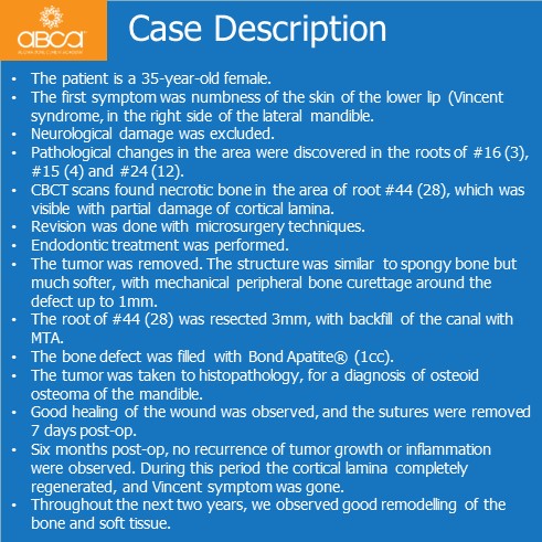
- The patient is a 35-year-old female.
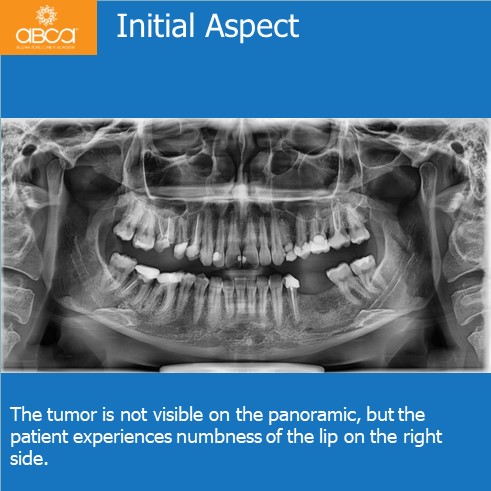
- The first symptom was numbness of the skin of the lower lip (Vincent syndrome, in the right side of the lateral mandible.
- Neurological damage was excluded.
- Pathological changes in the area were discovered in the roots of #16 (3), #15 (4) and #24 (12).
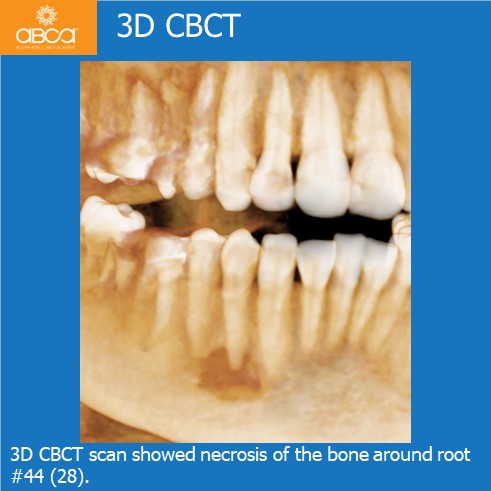
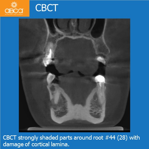
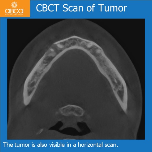
- CBCT scans found necrotic bone in the area of root #44 (28), which was visible with partial damage of cortical lamina.
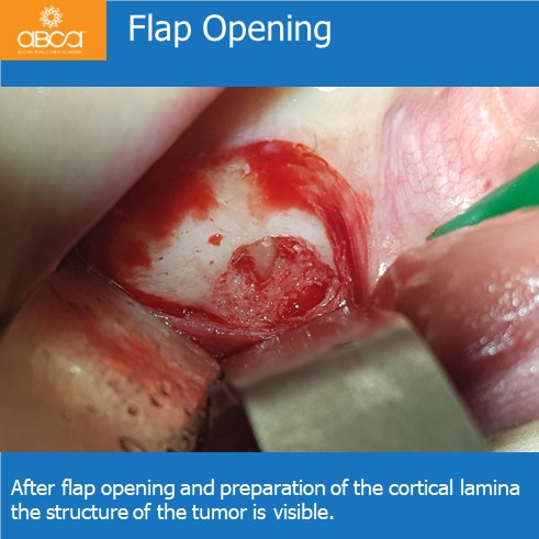
- Revision was done with microsurgery techniques.
- Endodontic treatment was performed.
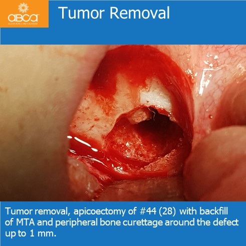
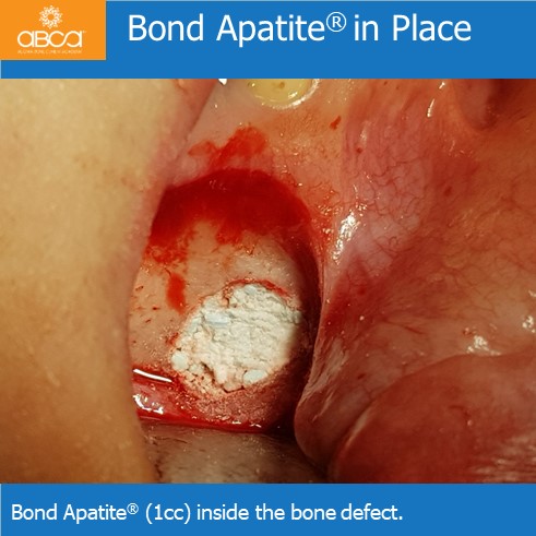
- The tumor was removed. The structure was similar to spongy bone but much softer, with mechanical peripheral bone curettage around the defect up to 1mm.
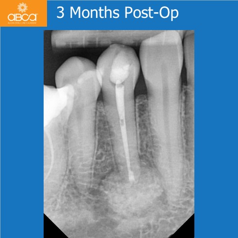
- The root of #44 (28) was resected 3mm, with backfill of the canal with MTA.
- The bone defect was filled with Bond Apatite® (1cc).
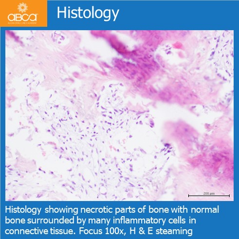
- The tumor was taken to histopathology, for a diagnosis of osteoid osteoma of the mandible.
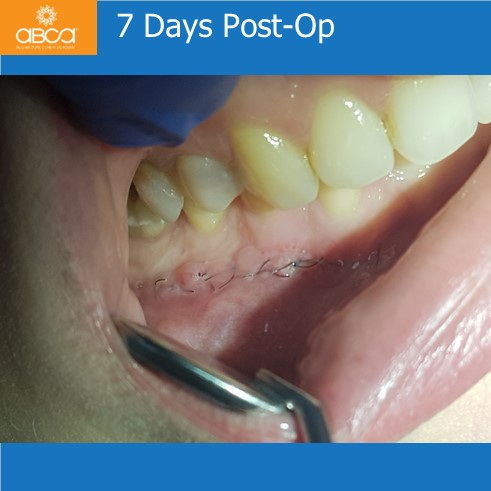
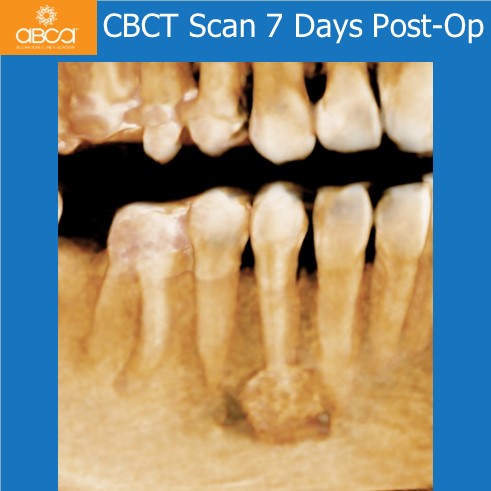
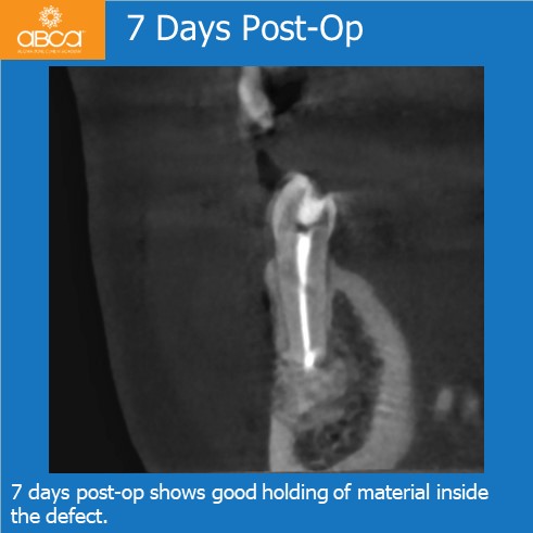
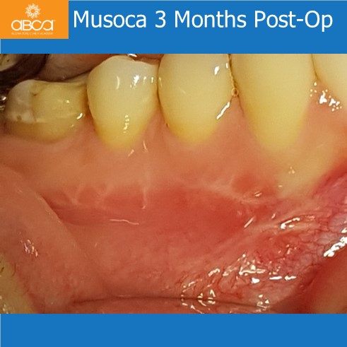
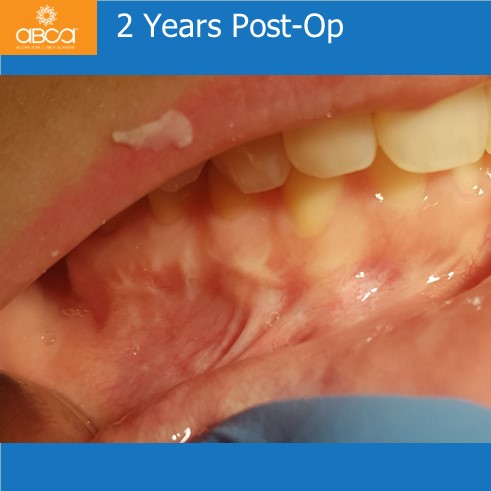
- Good healing of the wound was observed, and the sutures were removed 7 days post-op.
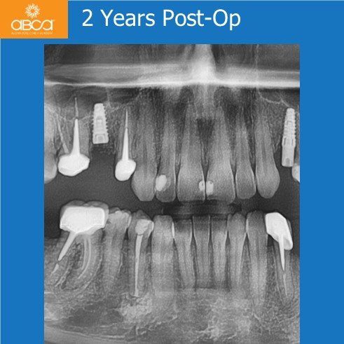
- Six months post-op, no recurrence of tumor growth or inflammation were observed. During this period the cortical lamina completely regenerated, and Vincent symptom was gone.
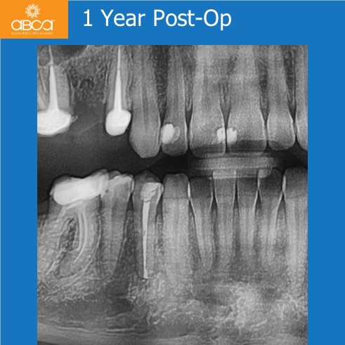
- Throughout the next two years, we observed good remodeling of the bone and soft tissue.